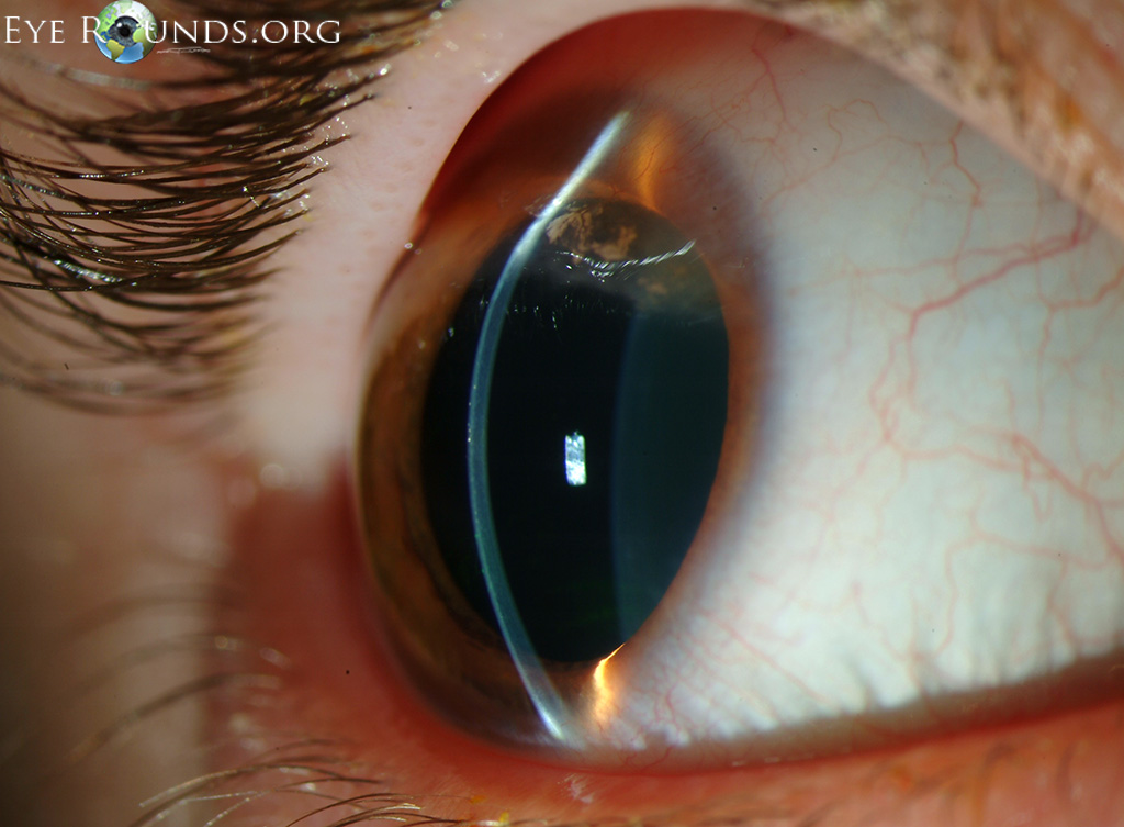


With these improved screenings, the known population with keratoconus has grown, and mild “forme fruste” cases that formerly escaped diagnosis are more readily detected. These measures are valuable for diagnosis because keratoconus often demonstrates abnormal anterior and posterior corneal curvatures in conjunction with corneal thinning.

Unlike corneal topography, which only measures the anterior cornea surface, Scheimpflug imaging and OCT can measure both the anterior and posterior corneal surfaces and perform pan-corneal pachymetry. Technologies such as corneal topography, Scheimpflug imaging and optical coherence tomography (OCT) have also helped identify more keratoconus patients ( Figure 1). Since the first FDA approval of an excimer laser for LASIK in 1999, increased screenings for LASIK candidacy have identified many patients who were previously unaware of their keratoconus. Nonetheless, the rise of LASIK has indirectly benefited those with keratoconus. Keratoconus is a well-recognized contraindication for undergoing LASIK because excimer ablation can further weaken and destabilize the keratoconic cornea, making vision much worse. This topographical map is typical for a keratoconus patient. ODs can make the most of the opportunity by updating their understanding of the disease and its many therapies.įig. Keratoconus is now an opportunity more than it is an obstacle. Today, Intacs (Addition Technologies) and corneal crosslinking (CXL) are new medical strategies, in addition to advanced hybrid and scleral contact lenses. Keratoconus was one of them, because management frequently involved eyeglass remakes, several contact lens prescriptions, rigid contact lenses that displaced and caused discomfort and, for the optometrist, historically inadequate third-party reimbursement.īut in the past two decades, several new options have turned this frustrating treatment paradigm on its head. The conditions ODs dread managing are those that highly impact patients’ lives but for which few treatments truly succeed. My Patient has Recurrent Corneal Erosion.Now What?Ĭorneal Manifestations of Systemic Diseases Check out the other feature articles in this month's issue:


 0 kommentar(er)
